Assessing Muscle Size with MuscleSound
Muscle Size is included in several Assessments, including
MuscleHealth Physical
Rehab
Muscle Size Assessment
How is Muscle Size calculated?
- MuscleSound identifies the muscle in the image
- The Centroid of the muscle is calculated in order to find the 'average middle'
- Muscle Thickness = (muscle centroid - fat muscle boundary) x 2
The timing of a scan is essential to provide a valid and reliable measure
MuscleSound determines the size of a muscle by measuring its thickness at predetermined locations. However, the fluid content of a muscle increases both during and following exercise. This will transiently increase the muscle volume and resulting in an erroneous assessment of its actual size.
The timing of scans for this measure needs to be considered for two separate, but linked situations
- A time frame for individual scan sessions
- A time frame for follow up scan sessions
Time frame for individual scan sessions
During exercise, blood and associated fluids shift into the muscle to meet the increased metabolic demand (Andersen & Saltin), a condition known as "Exercise-induced hyperemia” (Wray et al., 2005). This process, also referred to as “Transient Hypertrophy”, increases the size of the muscle, both during, and for a variable period of time after exercise. While the magnitude and extent of this well-established phenomenon are generally dependent on the mode, duration and intensity of the exercise session, there are wide individual variations in responses (Garton et al, 2014).
Optimal Times to measure Muscle Size
Muscle Size should be measured when the subject is rested and/or fully recovered from exercise
Measures are unlikely to be influenced by fluid shifts
Resting measures are the best reflection of a ‘normal’ status for muscle size
Similar conditions are required for other physiological measures such as blood pressure and pulse rate
Sub-Optimal times to measure Muscle Size
Muscle Size should not be measured within 2-3 hours after exercise
If scans are conducted too soon after an exercise session, the subject will not be fully recovered
Muscle Size is likely to be transiently increased as a result of exercise-induced hyperemia
Depending on the mode, duration and intensity, this transient increase in size can last up to 3 hours after cessation of exercise
Time frame for follow-up scan sessions
Over the last several decades, strength Increases occurring early in a training program were thought to be caused by neural adaptations in the muscle over a period of between 6-12 weeks. An increase in muscle size (Hypertrophy) was considered to be a relatively slow process, beginning only after these neural adaptations were completed (Moritani & Devries, 1979; Sale, 1988). However, recent studies using ultrasound have shown that hypertrophy can occur within the first 2-3 weeks of training. (Loenneke, et al., 2017; Seynnes, et al., 2007; Ogasawara et al., 2012; Krentz & Farthing, 2012). The magnitude of such changes and the capacity of ultrasound to detect them will vary according to the mode, intensity, and duration of exercise.
Optimal Times for follow up measurement of Muscle Size
Changes in Muscle Size should be monitored/measured after a minimum of 2-3 weeks of training
Research has shown that ultrasound is capable of detecting changes in Muscle Size over this time frame, depending on the mode of exercise, and assuming it is of sufficient duration and intensity.
Follow up scans can be repeated at similar intervals
Increases in Muscle Size will vary depending on the mode, intensity and duration of training
Sub-Optimal times for follow up measurement of Muscle Size
Changes in Muscle Size should not be monitored/measured within 2 weeks of training
Research has shown that ultrasound is unlikely to detect changes in Muscle Size within the first 2 weeks of a training program, regardless of mode, duration and intensity of exercise
Any changes detected within this period are likely to be produced by scanning errors or other artifact
What Muscles can be Assessed?
Currently the Muscles able to be Assessed are
- Thigh (Rectus Femoris - ‘RF’)
- Thigh (Vastus Lateralis - ‘VL’)
Future Muscles able to be assessed will include
- Upper Arm (Biceps Brachii - ‘BB’)
- Back of Thigh (Hamstring - ‘HL’)
- Calf (Gastrocnemius - ‘GS’)
How well does MuscleSound Measure Muscle Size?
Overview
Ultrasound provides a direct measurement of muscle thickness. This produces valid and reliable results (Tillquist, et al., 2014, Sanada, et al., 2006), as long as “Standard Procedures” are followed.
Ensuring Valid and Reliable Assessments of Muscle Size
This will ensure highly accurate and reliable measures
Standard error of measure = 0.66mm (Less than 1 mm)
Minimum Important Change, defined as "The smallest change detected that would be considered important/meaningful" = 1.8mm
For consistent results, the assessment must be taken pre-exercise when the client is fully rested and recovered. THIS IS CRUCIAL!
The scanned thickness of a muscle can transiently (and variably) increase both during and post exercise.
After High Intensity exercise, scanned muscle size can increase by as much as 25% compared to pre-exercise levels.
Probe is located at predetermined positions on the muscle. For example the midpoint of RF is standardized based on the height of the person being scanned.
Changing the location of probe by as little as +/- 1 inch can result in a muscle size change of up to +/- 12%.
Individual must be lying down relaxed on a flat and even surface (use a training table, or yoga mat).
Sitting up can result in a muscle size change of up to +/- 9% between these two body positions.
Uneven surface such as lying across a few chairs can result in a muscle size change of up to +/-8%.
NOTE: "Resting" the probe on the muscle is “Standard Protocol”.
Pressing the probe into the muscle will compress the tissues, resulting in an underestimation of muscle size.
Lifting the probe from the muscle tissues will allow them to expand, resulting in an over estimation of muscle size.
NOTE: It is possible to affect the muscle size by as much as 47% with incorrect probe pressure.
Bottom of the muscle must be visible on the image.
Increase depth of scan if necessary.
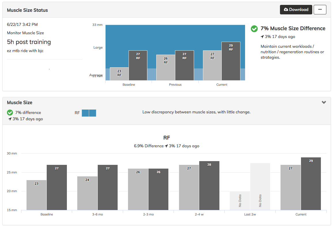
How are the Results Reported?
As Muscle Thickness (Size)
- Displayed in mm
As Percent Difference (Symmetry)
- Right vs Left
- See article on Muscle Size Symmetry
As Change from Baseline
- Trend, and amount of change
As Change from Previous Session(s)
- Trend, and amount of change
As Overall Status
- Based on percent difference and trend
Example of Reported Results
Comparison of Epidemiological and MuscleSound Data
Research has shown that Muscle Thickness (Size) can be considered a surrogate for Muscle Mass/Volume, particularly in the lower limbs (Abe, et al., 2015., Abe, et al., 2016., Ogawa, et al., 2012).
The two graphics in the left hand column below are taken from a study (Janssen, et al., 2000), published in the Journal of Applied Physiology that reports on skeletal muscle mass changes by age and gender. The two graphics in the right hand column below are taken from MuscleSound data on thigh muscle size collected over the last 4 years.
These data show that
- Age related loss of Total Muscle Mass closely patterns MuscleSound data on age related Muscle Size loss.
- The pattern of loss is even more evident between Thigh Muscle Mass and Rectus Femoris Muscle Size
|
|
|
|
|
|
OBSERVATIONS - Muscle Mass
|
OBSERVATIONS - Muscle Size
|


References
Abe, T., et al. Morphological and functional relationships with ultrasound measured muscle thickness of the lower extremity: a brief review. Ultrasound 23: 166–173, 2015
Abe, T., et al. Ultrasound assessment of hamstring muscle size using posterior thigh muscle thickness Clin Physiol Funct Imaging 36: 206–210, 2016
Andersen & Saltin. Maximal perfusion of skeletal muscle in man. J Physiol 366: 233–249, 1985.
Elsner & Carlson. Postexercise hyperemia in trained and untrained subjects. J Appl Physiol. 17:436-440, 1962.
Garten, et al. The role of muscle mass in exercise-induced hyperemia. J Appl Physiol. 116: 1204–1209, 2014.
Janssen, I., et al. Skeletal muscle mass and distribution in 468 men and women aged 18-88yr J Appl Physiol 89: 81–88, 2000.
Krentz & Farthing. Neural and morphological changes in response to a 20-day intense eccentric training protocol. Eur J Appl Physiol. 110:333–340, 2010.
Loenneke, et al. Time-course of muscle growth, and its relationship with muscle strength in both young and older women. Geriatr Gerontol Int. 1-8, 2017.
Moritani & deVries. Neural factors verses hypertrophy in the time course of muscle strength gain. Am. J. Phys. Med. 58: 115-130, 1979.
Ogasawara, et al., Time course for arm and chest muscle thickness changes following bench press training. Interventional Medicine & Applied Science. 4: 217–220, 2012
Ogawa, M., et al. Ultrasound Assessment of Adductor Muscle Size Using Muscle Thickness of the Thigh. Journal of Sport Rehabilitation 21: 244-248, 2012
Sale, D. Neural adaptations to resistance training. Med. Sci Sports Exerc. 20: S135-S145, 1988.
Sanada, K., et al. Prediction and validation of total and regional skeletal muscle mass by ultrasound in Japanese adults. Eur J Appl Physiol. 96: 24–31, 2006.
Seynnes, et al. Early skeletal muscle hypertrophy and architectural changes in response to high-intensity resistance training. Appl. Physiol. 102: 368-373, 2007.
Tillquist, M, et al. Bedside Ultrasound Is a Practical and Reliable Measurement Tool for Assessing Quadriceps Muscle Layer Thickness. J Parenter Enteral Nutr. 38:886-890, 2014.
Wray et al., Onset exercise hyperaemia in humans: partitioning the contributors. J Physiol 565: 1053–1060, 2005.